Thursday, March 12, 2015

Amblyopia in children

Children with moderate or severe amblyopia saw significant improvement in visual acuity with the use of daily 2-hour patching of the stronger eye, according to data collected in a trial's run-in phase published online February 19 in JAMA Ophthalmology.


psychological support also needed.

Friday, January 13, 2012

Proper arranged work station

it is so simple,
look at the picture

it is not only good for eye but also for back, neck, hip, leg and foot pains

Is it helpful ? Sunglasses for Health of Eyes


Sunglasses  protect our eyes from sunlight, as bright light is harmful for eyes.
While purchasing sunglasses don’t remember their actual purpose and do check the ability of glass against UV factor. Sunglasses protect your sensitive eyes from all sorts of bright lights.
Sunglasses are most important tool to guard you against glares while driving. Now it’s recommended to wear sunglasses while driving at day. As it reduce the risk of accidents by improving your vision. Glares are the reflection of sunlight when strikes metal or glass.Otherimportant role of sunglassesis the style statement. A wide range of glasses are available everywhere. Choose accordingly to your face cut, style, personality and age.

As we all know multi-storey buildings and vehicles are made of metals which strongly reflect back light making you unable to see for a moment. Protection of the eyes provided by sunglasses helps you to see clearly and prevents headaches. Special sunglasses are available which reduce glare affect for those who drive mostly. So hurry to buy sunglasses as they minimize the change of accidents due to glare.

Advantages of (GPL) Gas Permeable Lenses


To find the right contact lenses for your eyes it  can be very confusing. There are many varieties out there so it is essential you do your research well and find the best type for your eyes.
One of the lesser well known types of lenses are the gas permeable lenses. These are the most durable of lenses and also come under the name of rigid gas permeable, or oxygen permeable lenses.
Quite often people confuse these lenses with the old fashioned ‘hard’ lenses. These are now obsolete, but the gas permeable have taken their place and are far more reliable. The reason for the confusion is due to the ridged feel of the lenses that make some feel it is a hard lens.


The main reason why these lenses are not the most common is due to the fact that you will have to adapt to the lens far more than you do with a softer lens. Soft lenses are almost instantly comfortable, whereas gas permeable are not. But with time, they become comfortable and ultimately become more reliable.

Sunday, November 20, 2011

New treatment for macular degeneration

 November 18, 2011 -- The U.S. Food and Drug Administration approved Eylea (aflibercept) to be used in treatment of patients with wet (neovascular) age-related macular degeneration (AMD), which is one of major causes of vision loss and blindness in ages 60 and older.

AMD gradually destroys a person’s sharp, central vision. It affects the macula, the part of the eye that allows people to see fine detail needed to do daily tasks such as reading and driving.

There are two forms of AMD, a wet form and a dry form. The wet form of AMD includes the growth of abnormal blood vessels. The blood vessels can leak fluid into the central part of the retina, also known as the macula. When fluid leaks into the macula, the macula thickens and vision loss occurs. An early symptom of wet AMD occurs when straight lines appear to be wavy.

“Eylea is an important new treatment option for adults with wet AMD,” said Edward Cox, M.D., M.P.H, director of the Office of Antimicrobial Products in FDA’s Center for Drug Evaluation and Research. “It is a potentially blinding disease and the availability of new treatment options is important.”

The safety and effectiveness of Eylea was evaluated in two clinical trials involving 2,412 adult patients. People in the study received either Eylea or Lucentis (ranibizumab injection). The primary endpoint in each study was a patient’s clearness of vision (visual acuity) after one year of treatment.

Eylea is injected into the eye either every four weeks or every eight weeks by an ophthalmologist. The studies showed that Eylea was as effective as Lucentis in maintaining or improving visual acuity.

The most commonly reported side effects in patients receiving Eylea included eye pain, blood at the injection site (conjunctival hemorrhage), the appearance of floating spots in a person’s vision (vitreous floaters), clouding of the eye lens (cataract), and an increase in eye pressure.

Eylea should not be used in those who have an active eye infection or active ocular inflammation. Eylea has not been studied in pregnant women, so the treatment should be used only in pregnant women if the potential benefits of the treatment outweigh any potential risks. Age related macular degeneration does not occur in children and Eylea has not been studied in children.

Other FDA-approved treatment options for wet AMD include: Visudyne (verteporfin for injection) approved in 2000, Macugen (pegaptanib sodium injection) approved in 2004, and Lucentis (ranibizumab injection) approved in 2006.

Eylea is marketed by Tarrytown

Tuesday, October 25, 2011

Diabetic Retinopathy changes


Diabetic Retinopathy
In the normal eye, the lens is clear and transparent, which focuses light on the retina to produce sharp image and clear picture.
What happens in Diabetic Retinopathy due to diabetes is that: the blood vessels in the retina develop tiny leaks then retina becomes wet and swollen leading to hazy vision in Diabetic Retinopathy, the blood vessels of the retina become abnormal.
Some cases may develop wrinkling and detachment in retina (DR)
this video shows these abnormal changes


Diagnosis of all this and DR are diagnosed by proper eye examination with help of OCT Machine (fundus cameras), Angiography Machines
Treatment Available: Laser Surgery is prefered.

Monday, October 24, 2011

Continual Monitoring of Fluorescein Angiography

 A system has been developed which enables continuous monitoring and recording of circulating fluorescein in the retinal blood vessels. This is achieved with minimal modification of a Zeiss fundus camera. A night vision television camera (Ikegami) is used with illumination provided by the standard observation light of the fundus camera. This method offers advantages over standard fluorescein angiography in patient comfort, information obtained and the immediate evaluation of the fluorescein angiograms being available.

Saturday, October 8, 2011

Checklist and preparation for fluorescein angiography


Checklist and preparation for fluorescein angiography:

1. Informed consent.

2. Inform the patient about fluorescein angiography. Obtain verbal or written informed consent.

3. Dilate patient pupil.

4. Prepare fluorescein solution, vein needle, and syringe.

5. Prepare fundus camera:
a-clean front lens.
b-load color and black-and-white film into two camera backs.
c-focus eyepiece crosshairs.

6. Take identification photograph, which includes the patient name, the date, and other data (number of fluorescein angiogiogram, patient vision of right and left eye, referring physician and so forth)

7. Position the patient for alignment,focus,and comfort.

Screening for Diabetic Retinopathy

Diabetes causes vasculopathy and retinopathy, so it needs good care and continuous follow up
screening for diabetic patients for retinopathy is done by Fundus Fluorescein Angiography.


diabetic patients should go for regular visits to ophthalmologist for early detection of retinopathy and monitoring its progression 


Fluorescein pathway and timing


Fluorescein pathway:
Arm-to-retina circulation time is 12-15 sec.
angiography dye injection
(Five angiographic phases):

  • Pre arterial
  • Arterial
  • Arteriovenous
  • Venous
  • Recirculation

Modes of retinal photography

1. Color, where the retina is illuminated by white light and examined in full color.
retinal color photography

2. Red-free, where the imaging light is filtered to remove red colors, improving contrast of vessels and other structures.

BENEFITS OF DIGITAL PHOTOGRAPHY


BENEFITS OF DIGITAL PHOTOGRAPHY:
  1. Instant knowledge of image quality
  2. Networking of images (a must)
  3. Reduction in administration time
  4. No film / development costs
  5. Easy export for presentations / e.mail
  6. Instantly available for viewing
  7. Comparison over time on screen
  8. Digital image manipulation tools
  9. Linkage to Electronic Patient Records
  10. Diagnostic Database

patients doesn’t need angiography

When the patients doesn’t need angiography:


  • AMD doesn’t need treatment : disciform scar, geographic atrophy , senile derusen , atrophic scar after PDT.
  • Choroidal melanoma.
  • Retinal vascular obstruction only in retinal vein occlusion after 3 months for laser treatment.
  • Congenital retinal dystrophy (fundus autofluoresence is preferable).

Causes of bad quality angiography


Causes of bad quality angiography :


• Speed of dye injection (2-5 sec)

• Ocular media opacities (cataract , vitreous hemorrhage , corneal scar)

• Patient movement (image out of focus)

• Dust on the camera.

• The film (out of date)

• Problem in print process.

• Insufficient Pupil dilatation (at least 6mm) specially with mydriatic cameras.

• Inadequate tear film.

Thursday, September 29, 2011

Fundus Fluorescein Angiography (FFA), eye angiogram

Fluorescence :
is a property of substance to alter the wave length of the reflected light on exciting. Angiography means recording of the angios or the blood vessels. It is basically recording and visualization of the blood vessels of the retina using fluorescein dye.

Characteristics of Fluorescein :
Nontoxic, inexpensive, safe
Alkaline solution
Highly fluorescent
Absorbs blue light (480-500 nm)
Emits yellow-green (500-600 nm [525 nm])
Effective at pH 7.37-7.45
Removal from blood by kidneys and liver within 24 hrs.

During Pregnancy and Lactation :
Controversial
Fl. Crosses the placenta
Has been done in pregnancy with no adverse effect
Do it when necessary
Fluorescein is secreted in milk.


fluorescein dye arm injection
General overview on Procedure :
In FFA, the chemical or dye is injected in the vein of the hand and this dye circulates all over the body and in every blood vessel. Since retina is the only place in the body where blood vessels can be seen, it is used to visualize the retinal vasculature, that is blood vessels of the retina.
A special machine called the fundus camera, which has got special lens filters, is used to take photographs of the retina. We can also attach digital camera system or a printer.
When blue light falls on retina which has Fluorescein dye the reflected light is of green colour & not blue. This property of dye is used to study the normal or abnormal anatomy of retina. This dye and the reflected light is greenish in color. The fundus camera records this. This dye would go wherever the blood vessels are present in retina and we would get the picture of that.

If blood vasculature of the retina is normal, then we would get the normal pattern of the retina. If there were blockage then the dye would not go beyond that and would stop at that particular point. If there is leakage, the dye would also leak out along with the blood in the retina and we would be able to pick up that. If there are certain other defects in the retina then depending on the various features seen, whether we get leakages or staining or blocked fluorescence or window defects whether the dye seen in early phase, late phase or in very late phase or delayed phase and is initially less and increasing etc, we can get very good idea of patients disease.

retinal angiography procedure

Tuesday, September 27, 2011

precautions with Diabetic Patients


precautions with Diabetic Patients :
Diabetic patients: this dye test gives false high readings in the urine and on blood sugar tests. You should not adjust your insulin or any of your diabetic treatments based on these results during the first 2 days following the Fluorescein angiogram. so

Sunday, September 25, 2011

Special precautions with diabetes, kidney diseases, high blood pressure or receiving anticoagulants


Special precautions to take before having an angiogram:

Patients with diabetes, kidney diseases, high blood pressure or receiving anticoagulants have to take special precautions as following and an angiography should only be done only when it is necessary.


 If you have kidney problems an angiography should only be done if clinically indicated as the dye is removed from the blood by the kidneys and can cause further impairment of kidney function.
 If you are diabetic patient: this dye test gives false high readings in the urine and on blood sugar tests. You should not adjust your insulin or any of your diabetic treatments based on these results during the first 2 days following the Fluorescein angiogram. 
* If you suffer from high blood pressure (hypertension) then this should be well controlled with tablets before angiography is done as there is a higher risk of bleeding from the puncture site.
* If you are receiving anticoagulant drugs such as WARFARIN then these should be stopped before angiography  is done and may need to be replaced with a more controllable drug such as HEPARIN.
* If you have a known allergy to fluoresceine then great care should be exercised in the use of fluoresceine dyes that are used in angiography.

Saturday, September 24, 2011

Side effects of fundus angiography


Safety of Fluorescein :
Topical, oral, and intravenous use of fluorescein can cause adverse reactions including nausea, vomiting, hives, acute hypotension, anaphylaxis and related anaphylactoid reaction, cardiac arrest, and sudden death. Intravenous use has the most reported adverse reactions, including sudden death, but this may reflect greater use rather than greater risk. Both oral and topical uses have been reported to cause anaphylaxis, including one case of anaphylaxis with cardiac arrest (resuscitated) following topical use in an eye drop. Reported rates of adverse reactions vary from 1% to 6% The higher rates may reflect study populations that include a higher percentage of persons with prior adverse reactions. The risk of an adverse reaction is 25 times higher if the person has had a prior adverse reaction. The risk can be reduced with prior (prophylactic) use of antihistamines and prompt emergency management of any ensuing anaphylaxis. A simple prick test may help to identify persons at greatest risk of adverse reaction.


Simple side effects:
  • Nausia
  • Vomiting
  • Some kind of pain in the hand
  • Yellow colored discoloration of urine
  • Blurring of vision and some dazzle from the camera’s flash
  • The dye can tint your skin a slight yellow

Management of simple hazards:
Most people do not experience any side effects apart from blurring of vision and some dazzle from the camera’s flash.

The nausea usually is transient and subsides quickly. Hives can range from a minor annoyance to severe.
A single dose of antihistamine may give complete relief.

The dye can tint your skin a slight yellow colour and your urine can

Friday, September 23, 2011

Fundus Cameras

Retinal Imaging:
The eyes are the window to the soul 16th century proverb
The retina gives an unobstructed view of the human vascular.
Also, retinal imaging is important for diagnosing retina diseases.


Fundus camera:
Is a device for photographing the retina.
It is based on the indirect ophthalmoscopy principle, where the Observer’s eye is replaced with a camera.

So, A fundus camera or (retinal camera)
Is a specialized low power Microscope with an attached Camera designed to Photograph the interior surface of the Eye, including the Retina, Optic disc, Macula, and Posterior pole i.e. the Fundus (eye).


Optical principles:
The optical design of fundus cameras is based on the principle of monocular indirect ophthalmoscopy.
A fundus camera provides an upright, magnified view of the fundus. A typical camera views 30 to 50 degrees of retinal area, with a magnification of 2.5x, and allows some modification of this relationship through zoom or auxiliary lenses from 15 degrees which provides 5x magnification to 140 degrees with a wide angle lens which minifies the image by half. The optics of a fundus camera are similar to those of an indirect ophthalmoscope in that the observation and illumination systems follow dissimilar paths.

The observation light is focused via a series of lenses through a doughnut shaped aperture, which then passes through a central aperture to form an annulus, before passing through the camera objective lens and through the cornea onto the retina.The light reflected from the retina passes through the un-illuminated hole in the doughnut formed by the illumination system. As the light paths of the two systems are independent, there are minimal reflections of the light source captured in the formed image. The image forming rays continue towards the low powered telescopic eyepiece.
When the button is pressed to take a picture, a mirror interrupts the path of the illumination system allow the light from the flash bulb to pass into the eye. Simultaneously, a mirror falls in front of the observation telescope, which redirects the light onto the capturing medium, whether it is film or a digital Charge-coupled device.

Thursday, September 22, 2011

Topcon retinal cameras (TRC-NW7SF MK II)


TRC-NW7SF MK II (Topcon)


Description:

The TRC-NW7SF offers you the versatility of dilated and undilated retinal imaging in both black & white and colour modes. In the non-mydriatic mode, the TRC-NW7SF can make colour, red-free and fluorescein images. In the mydriatic mode, it can also shoot ICG live and still images.

For ease of alignment and focusing, 6.4 inches colored monitor is available. This monitor can be used for capturing still and live images and with its ability to be angled a full 90 degrees side to side, allows the operator to concentrate on getting the patient perfectly positioned. TRC-NW7SF has a touch screen control panel for simple changes in procedures. Each procedure has its own pre-setting and background colour. The touch screen incorporates smart features such as small pupil mode and aperture adjustment for an enhanced depth of field. The swing and tilt mechanism allows viewing of peripheral segments without moving the patient’s head or fixation.

Key Features

-The ultimate all-in-one OCT.
-High resolution OCT.
-High resolution colour fundus image.
-High resolution FA & FAF images.
-Intuitive workflow.
-Normative database.
-Glaucoma module.
-network support (Full).
-IMAGEnet i-base connection.


specifications:


Tuesday, August 30, 2011

NIDEK (Non-Mydriatic) Auto Fundus Camera

NIDEK delivers the innovative non-mydriatic digital fundus camera that integrates every function required for easy retinal screening. Customized built-in functions of the NIDEK AFC-230/210 improve the quality and efficiency of medical examinations.
It has a screen connection.


Features
  • Innovative and unique Auto Focus and Auto 3D* Tracking.
    *AFC-230 Only
  • High Quality Retinal Imaging
    Integrating the innovative imaging optical system, this technologically advanced AFC-230/210 realizes digital fundus imaging of high resolution and fine gradation.
  • Accurate Anterior Eye Observation before Photography
    The AFC230/-210 integrates the 5.7-inch TFT LCD (640 x 480) monitor in addition to the special optic system.
  • Unique Blink Control
    With the automatic blink detection, the AFC-230/210 automatically stops the photography when the patient blinks.
  • Anterior Eye Photography Mode
  • Smaller Pupil Diameter Mode
    In addition to the regular minimum Pupil Diameter ø4.0 mm, the AFC-230/210 is also highly capable of detecting a smaller Pupil Diameter Minimum 3.7 mm.
  • Stereo Mode*
    Stereo fundus photography is also possible.
    *Requires stereo viewer (optional).

Kowa (nonmydriatic / mydriatic) fundus camera


fundus camera


VX-10α

Three photographic modes

*3 modes with one-touch switch ( nonmydriatic / mydriatic / fluorescein ).

*Mydriatic color and fluorescein photography can now be aligned through the LCD observation monitor.
*multiple step flash for proper exposure in various photographic modes.

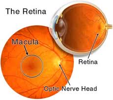
*Built-in internal fixation target for optic nerve head photography.
*Built-in small pupil mode (during mydriatic color and fluorescein photography) for patients with insufficient pupillary dilation


Easy functionality
Easy alignment and focusing adjustment with the LCD observation monitor.
Operationally focused, darkroom adapted navigation panel.
Clear viewfinder with a Long Eye Relief design.
Simple focusing with the point matching method.
Electric insertion of the fluorescein filter.


Adapted to the newly developed CCD camera

High resolution images when combined with the 2.1 megapixel digital CCD camera (KD-211C)(1600X1200 pixels image size) or the 6 megapixel digital CCD camera.
VX-10α combined with the VK-2 imaging system is part of an ideal digital imaging environment with multiple digital/video image inputs and networking

For advanced digital SLR imaging


Newly developed video adapter enables you to take both standard color photography and fluorescein angiography by high resolution Nikon* digital SLR camera.
All taken images are transferred the VK-2 imaging system automatically.


Options

Video adapter (for KD-211C)
Video adapter (1/2" bayonet)
Video adapter (for specific Nikon* digital SLR camera)
2.1 megapixel digital CCD camera KD-211C
Green (red free) filter
Grip
KOWA VK-2 Digital imaging system

Canon Retinal Cameras


Retinal Camera


Canon CX-1 Retinal Camera


The CX-1 is a MYD and Non-MYD hybrid digital retinal camera. It is extremely versatile with its 5 photography modes; colour. FAG, red free, cobalt and FAF (Fundus Auto Fluorescence). Making it ideal for screening and the diagnosis of the main eyes diseases.

Features

  • Digital Retinal Camera,
  • Hybrid camera MYD / Non MYD
  • FAF (Fundus Auto Fluorescence), also in Non Myd mode
  • Use of EOS technology
  • Advanced Stereo photography system
  • Bundled Retinal Imaging Control Software
  • Open connectivity and DICOM compliant
Canon’s CX-1 is a compact and portable hybrid retinal camera that combines both Mydriatic and Non-Mydriatic Modes, and switches between the two with a simple touch of a button.


Hybrid camera
:
Canon’s CX-1 is a compact and portable hybrid retinal camera that combines both Mydriatic and Non-Mydriatic Modes, As well as saving on equipment investment, this function also means that the examiners using the equipment can screen for more than one disease, e.g. AMD (Age-related Macular Degeneration), Glaucoma and diabetic retinopathy, with the same unit. This saves time and removes the need for repeat appointments.

5 Photography Modes
With the CX-1 following photography modes are available; Color, Red-free, Cobalt, Fluorescein Angiography and Fundus Autofluorescence (FAF) Alignment and observation with the CX-1 can be done through the viewfinder or by the large EOS LCD screen

FAF (Fundus Auto Fluorescence)
Detecting Auto fluorescence of the retina is an important indication of the retina’s health. With the CX-1 it is possible to take FAF (Fundus Auto fluorescence) images, even in Non-Mydriatic mode.

Use of EOS Technology
Canon’s own EOS camera technology, with its renowned image processing capabilities, is adapted exclusively for medical use in the CX-1 to provide optimal retinal imaging in a compact and convenient system. The single onboard 15.1 MegaPixel digital camera handles five different photography modes with ease, including non-mydriatic FAF photography, allowing EOS imaging technology to benefit all retinal images from the CX-1.

Enhanced stereo photography function
Easy capture for stereo view, by using the stereo guide marks on EOS LCD in the non-mydriatic mode, or stereo unit (option) a stereo pair can be created and managed very simply

Bundled Retinal Imaging Control Software
Bundled Retinal Imaging Control Software (RICS) for full camera control and image optimalization. The software has extensive diagnostic tools for optimized workflow and patient management; e.g. Cup/Disc ratio calculation, compare studies, RGB channel display, stereo viewing

Open connectivity and DICOM compliant
The CX-1 with RICS has open connectivity with existing networks and is fully DICOM compliant.